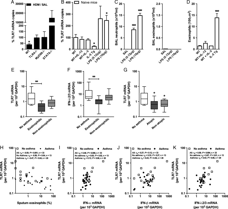
Figure 4.
Toll-like receptor (TLR)7 expression is reduced during eosinophilic lung inflammation. (A) Wildtype (WT), TLR4−/−, MyD88−/− and Stat6−/− mice were sensitised and challenged with house dust mite (HDM) over 18 days and gene expression of TLR7 in lower airway tissue was quantified by quantitative RT-PCR. TLR7 airway expression in allergic airways as a percentage of sterile saline (SAL) expression for each strain, (B) as well as lung tissue eosinophils and (C) bronchoalveolar lavage (BAL) fluid neutrophils and eosinophils (D) from rIL-5, rIL-13, IL-5 Tg and lipopolysaccharide (LPS)-treated mice 24 h post treatment. Results are mean±SEM (n=4–6 mice per group), gene expression in mice expressed as a % compared to non-allergic SAL-treated wildtype mice. TLR7 (E) and interferon (IFN)λ2/3 (F) expression in bronchial biopsies collected from non-asthmatic (n=13) and asthmatic (n=20) subjects stratified into eosinophilic (>3% eosinophils) or non-eosinophilic (<3% eosinophils) phenotypes based on BAL cell counts. (G) TLR7 expression in bronchial biopsies collected from non-asthmatic (n=13) and asthmatic (n=20) subjects stratified according to atopic status. Lines indicate the median, boxes extend from 25th to the 75th percentile, and error bars extend to 10th and 90th percentiles. (H) Correlation between TLR7 expression from bronchial biopsies and percentage of sputum eosinophils. (I–K) Correlations between TLR7 and IFN expression from patient biopsies. *p<0.05, **p<0.01, ***p<0.001 as determined by Student's t test (A–D) or Kruskal–Wallis with Dunn's multiple comparisons test.

