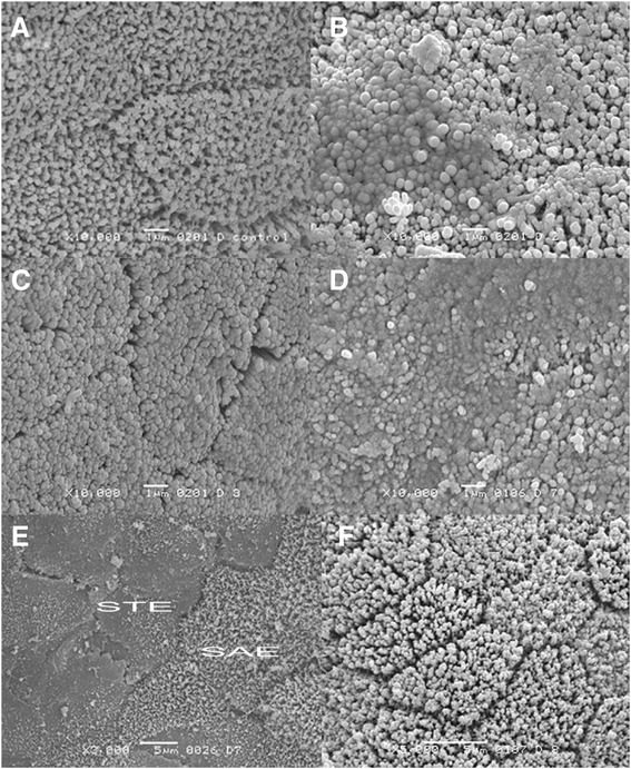
Fig. 2.

Scanning electron micrographs of rabbit duodenal mucosa, showing ultraestructural changes in enterocytes. a Villi of normal duodenal mucosa (scale bar = 1 μm, original magnification 10000x). b Villi of animals treated with acetylsalicylic acid for 14 days, with microvilli fused (scale bar = 1 μm, original magnification 10000x). c Villi of rabbits treated with Plantago ovata husk + acetylsalicylic acid for 14 days, with clearly defined limits in enterocytes (scale bar = 1 μm, original magnification 10000x). d Villi of animals treated with acetylsalicylic acid for 28 days, with microvilli aggregated (scale bar = 1 μm, original magnification 10000x). e Villi of animals treated with acetylsalicylic acid for 28 days, with slightly affected enterocytes (SAE) and strongly affected cells (STE). When several strongly affected enterocytes were adjacent, it is not possible to delimitate cells (scale bar = 5 μm, original magnification 2000x). f Villi of rabbits treated with Plantago ovata husk + acetylsalicylic acid for 28 days. Cell limits are well defined and exhibited no changes (scale bar = 5 μm, original magnification 5000x)
