
Figure 1.
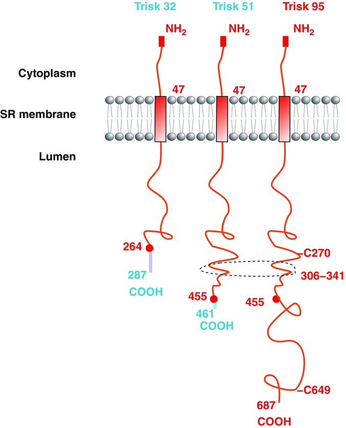
Structure of the main rat triadin isoforms The structures of the triadin isoforms in the sarcoplasmic reticulum are identical: a common N-terminal domain in the cytoplasm, a transmembrane helix, and a luminal part of variable length with a unique C-terminal end. The common parts are in red and the specific segments, starting at the red circles corresponding to the last common amino acids, are in a different colour. The domains involved in membrane deformation and indirect interaction with the microtubules are shown: the two cysteines C270 and C649, and the coiled-coil segment 306–341. The numbers refer to the rat sequence.