
Figure 6.
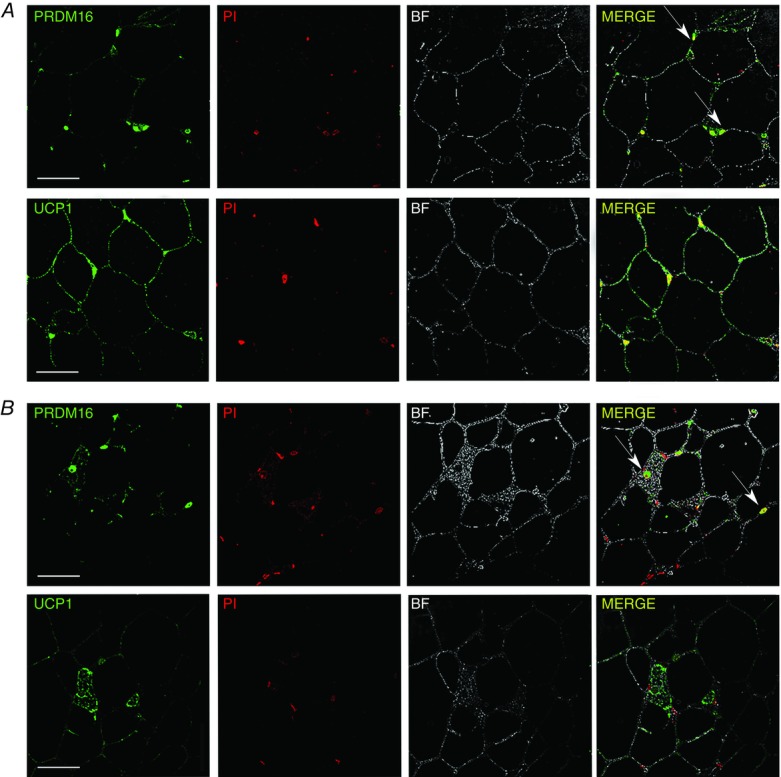
Immunofluorescence of UCP1 and PRDM16 in rpWAT of rats maintained at 4 ± 1°C for 3 (A) or 45 days (B)
Immunofluorescence labelling of unilocular/paucilocular and multilocular adipocytes for UCP1 (green) showing all adipocytes in rpWAT pass through the brown-adipocyte-like molecular stage during the early stages of cold acclimation, as demonstrated by the elevated UCP1 levels (A). After cold acclimation, however (B), only a small number of multilocular adipocytes exhibit brown-like appearance and were immunopositive for UCP1. PRDM16 protein levels do not directly correlate with the recruitment status of rpWAT adipocytes. PRDM16-immunopositive nuclei are visible in rpWAT adipocytes after 3 (A) and 45 (B) days of cold acclimation regardless of adipocyte appearance. Furthermore, the PRDM16 signal was diminished in multilocular (UCP1-expressing) adipocytes compared with adjacent unilocular/paucilocular (UCP1-negative) adipocytes after a 45-day cold acclimation (B). PRDM16 immunofluorescence (green) and staining of cell nuclei with propidium iodide (red) resulted in PRDM16 appearing yellow in the merged image. Nuclei of PRDM16-positive adipocytes are marked with arrows. UCP1 and PRDM16 were not stained and therefore absent in control samples (not shown). Scale bars = 50 μm.