Figure 3.
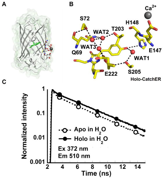
Structure and fluorescence lifetime of CatchER (A) The X-ray crystal structure of Ca2+-CatchER (PDB ID: 4L12). The residues shown in stick are the designed calcium binding site. Residues 164-168 were shown with 50% transparency. The residue shown in green is the chromophore. The atom shown in grey is the calcium atom. The oxygen atoms and nitrogen atoms are indicated in red and blue, respectively. (B) The proton wire observed in the crystal structure of Ca2+-CatchER (PDB ID: 4L12). The H-bonds are shown in the black dash line with the cut off of 3.5. Only side chains were shown in S72, T203, S205 and E222. (C) Fluorescence decay traces of apo-CatchER (dash line) and CatchER supplemented with 10 mM Ca2+ (Holo, solid line). CatchER was excited at 372 nm and emitted at 510 nm in the time range of 15 ns. Ex and Em are short for excitation and emission, respectively. Reprinted (adapted) with permission from Zou, et al., J. Phys. Chem. B, 2014. Copyright © 2015 American Chemical Society.

