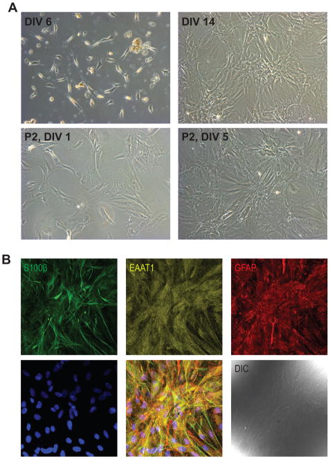
Figure 1.
A. Representative examples of ONHA culture after 6 days in vitro (DIV) prior to the first media change. Note the attached ONHAs and remaining tissue debris from the dissociation. The same culture at DIV14 after two media changes. ONHA passage 2 imaged 24 hr after seeding into a new tissue culture flask at a 1:5 dilution and the same culture, 5 days after seeding. B. ONHAs showed strong positive immunoreactivity for the astrocyte markers S100β, EAAT1, and GFAP. Representative, single confocal section is shown. Cells were from passage 5. Scale bar: 100 μm.

