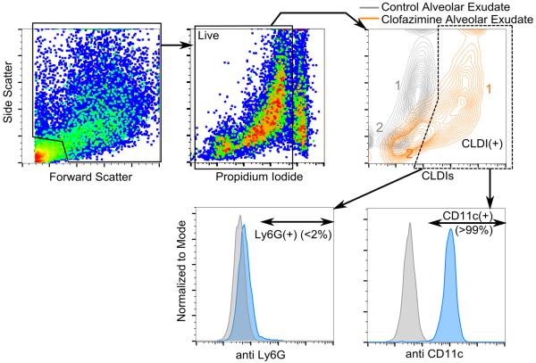
Figure 6.
Flow cytometric analysis of alveolar exudate at 8 weeks post drug feeding. Propidium iodide (PI) fluorescence was detected using the compensated 561 614/20 laser-detector setting where CLDI fluorescence was detected using the 640 671/30 setting. Gating was done on live cells followed by CLDI(+) cells. The population labeled 2 in orange color showed significant CLDI signal marking them out as CLDI(+) cells. The alveolar exudate was also labeled with antibodies for the macrophage surface marker CD11c (detected using 405 448/59) or the neutrophil surface marker Ly6G (detected using 405 448/59). CLDI(+) cells were overwhelmingly CD11c(+) (>99%, 6 pooled mice lavage sample, repeated twice) and Ly6G(−) (6 pooled mice lavage sample, repeated twice). Gating for antibody positivity was done using the corresponding isotype (grey background histogram).

