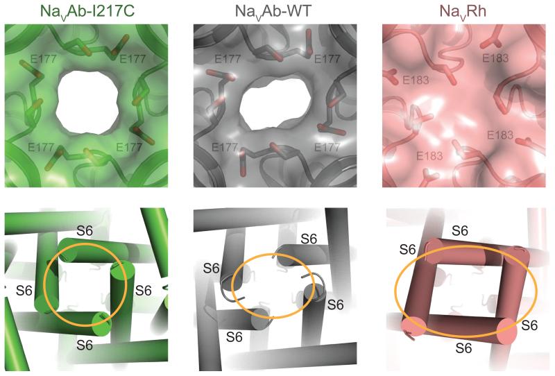
Figure 4. Structural Basis of Slow Inactivation.
Specific structural features of NaVAb-WT and NaVRh, including a distorted selectivity filter and the breakdown of the four-fold symmetry of the pore, as expected for the slow inactivation state. Top panel: Close-up views of the extracellular entrance of NaVAb-I217C (green, symmetric), NaVAb-WT (grey, asymmetric), and NaVRh (salmon, collapsed) with semi-transparent surface representation of the three channels [9-11]. The high-strength-field site glutamate residue in each structure along with its nearby serine residue is shown in sticks. Bottom panel: Close-up view of the intracellular closed activation gate of NaVAb-I217C (symmetric, orange circle), NaVAb-WT (asymmetric, orange oval), and NaVRh (asymmetric, orange oval). .

