
Figure 1. BFP assay and control experiments.
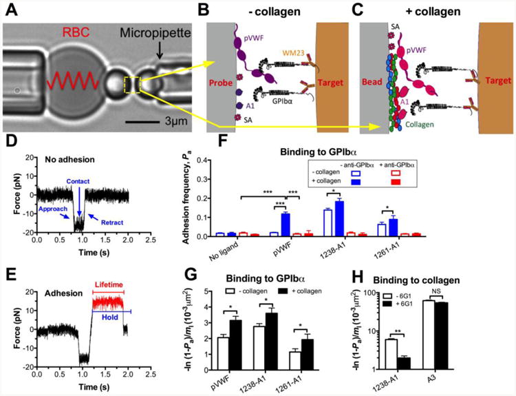
(A) BFP photomicrograph A micropipette-aspirated RBC with a bead (left, termed “probe”) attached to the apex formed a pico-force sensor, as depicted by a spring. It was aligned with another bead (right, termed “target”) aspirated by an apposing micropipette. (B and C) BFP functionalization. VWF-A1 or plasma VWF (pVWF) was coupled to the probe bead (left) via either direct linking to a plain surface, denote “- collagen” (B) or indirect linking to a collagen-bound surface, denote “+ collagen” (C). GPIb-IX was captured from platelet lysates by antibody WM23 covalently precoated on the target bead (right). Streptavidin (SA) was also covalently coupled to probe beads for attachment to biotinylated RBCs. (D and E) Force vs. time traces from two representative test cycles. A target bead was driven to approach a probe bead (∼0 pN), contacted for certain duration, retracted and ended the cycle if no adhesion (D) or held at a preset force (12 pN) until dissociation (signified by a force drop to zero) if adhesion was detected (indicated in red) (E), the duration of which was measured as bond lifetime (∼0.6 s). (F) Binding specificity. Adhesion frequencies (Pa) between the GPIbα targets and probes coated without or with indicated VWF ligands in the absence (blue) or presence (red) of 50 μg/ml anti-GPIbα (AK2) blocking mAb. (G) Average number of A1–GPIbα bonds per unit A1 site density [-ln(1 - Pa)/m1], which equals to the product of effective 2D affinity (AcKa) and GPIbα site density (mr). The same mr was used to test all VWF ligands. The results from “- collagen” (open) and “+ collagen” (closed) preparations are compared. (H) Collagen binding. Adhesion frequencies between the collagen III target and probes coated with 1238-A1 or A3 in the absence (open) or presence (closed) of 50 μg/ml anti-A1 (6G1) mAb. Each probe–target pair was tested repeatedly for 100 approach-contact-retract cycles to estimate a Pa. Five probe–target pairs were tested to obtain mean ± S.E.M. NS = not significant; * = p < 0.05; *** = p < 0.001, assessed by unpaired, two-tailed Student's t-test.