
Figure 4. Characterization of collagen binding induced A1 conformational change by antibodies.
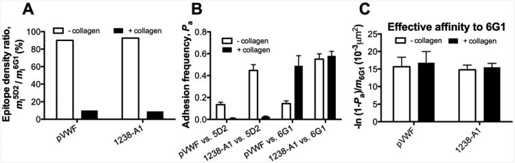
(A) Normalized A1 site densities detected by conformation specific anti-A1 mAbs. The ligand site densities for pVWF and A1 were measured by flow cytometry using 5D2 (m15D2), then normalized by the site densities measured with conformation-independent 6G1 (m16G1). The data were presented as m15D2/m16G1 in percentage. (B) Conformations probed by mAb coated targets. Adhesion frequencies (Pa) between the 5D2 or 6G1 coated target and probes coated with A1 or pVWF. (C) The effective binding affinities of pVWF and 1238-A1 to 6G1 by converting Pa6G1 (from panel B) to [-ln(1 - Pa6G1)/ m16G1]. The same keys were used as Fig. 1G.