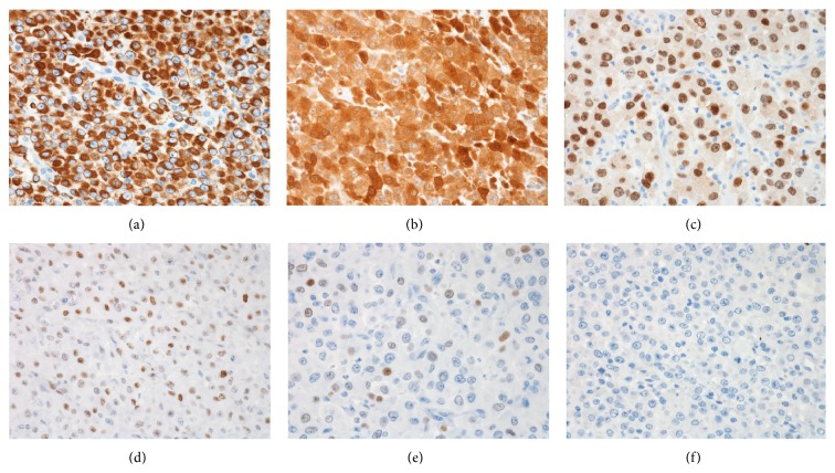
Figure 3.
Immunohistochemical features of the tumor cells. The tumor cells showed diffuse positivity for AE1/AE3 (a), galectin-3 (b), and PAX-8 (c). There was patchy positivity for p53 (d). The tumor cells showed only weak focal positivity for TTF-1 (e) and were negative for thyroglobulin (f). All photographs at 400x magnification.

