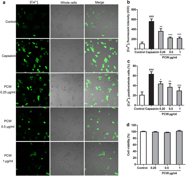
Fig. 7.

Effect of PCW on capsaicin-induced [Ca2+]i influx in HEK293T-RPV1 cells. HEK293T-RPV1 cells were incubated with PCW (0.25, 0.5 and 1 μg/ml, respectively) for 30 min, then Fluo-4AM for 1 h, and immediately stimulated with or without capsaicin (8 µM). The cells were divided into five groups: Control–HEK293T-TRPV1 cultured cells; Capsaicin–capsaicin-induced cells; PCW groups–capsaicin-induced cells treated with various concentrations of PCW (0.25, 0.5 and 1 μg/ml, respectively). a Localization of [Ca2+]i (green) in HEK293T-RPV1 cells with by fluorescence staining and laser scanning microscopy. b Fluorescent intensity of [Ca2+]i in HEK293T-RPV1 cells. c Mean change in fluorescence ratio in HEK293T-RPV1 cells. 10 microscopic fields were selected randomly and [Ca2+]i positive cells were counted. d No effect of PCW (0.25, 0.5 and 1 μg/ml, respectively) on the cell viability by 3-(4,5-Dimethylthiazol-2-yl)-2,5-diphenyltetrazolium bromide method. Cell viability of the control was taken as 100 %. Data are represented as the mean ± SEM. ### P < 0.001 vs. the control group; *P < 0.05, **P < 0.01 and ***P < 0.001 vs. the capsaicin group, respectively. n = 3 in each group and each assay was repeated 2 times
