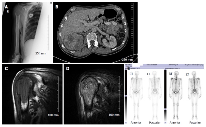
Figure 4.

Lytic bone metastases are poorly demonstrated on bone scintigraphy. Plain radiograph (A) demonstrating a lytic metastatic deposit in the right proximal humerus in a patient with a large right renal cell carcinoma (B); Corresponding abnormal low T1 and high short tau inversion recovery signal on magnetic resonance imaging (C and D); Only the small osteoblastic component of the metastatic deposit demonstrates abnormal accumulation of radiotracer on bone scintigraphy (E).
