Figure 6.

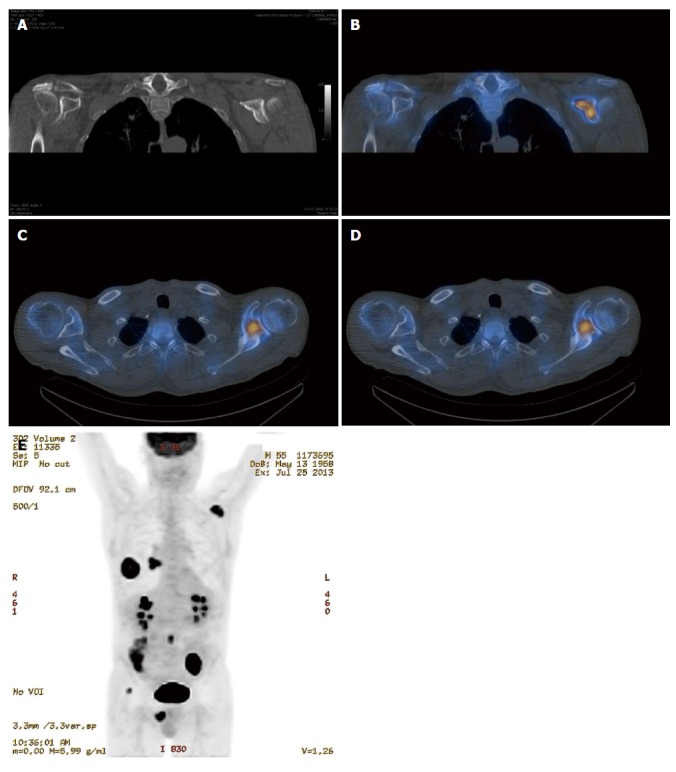
Single photon emission emission computed tomography-computed tomography is more sensitive for detection of bone metastasis than computed tomography alone. A: Coronal CT image of the left scapula (bone window) in a patient with primary lung malignancy does not demonstrate an aggressive bone lesion; Coronal and axial single photon emission CT/CT (B, C) and axial 18F fluorodeoxyglucose-positron emission tomography (FDG-PET)/CT (D) demonstrate abnormal radiotracer accumulation in the left clavicle consistent with bone metastasis; E: Coronal PET maximum intensity projection image demonstrating 18F FDG avid primary lung malignancy and right hilar lymph node metastasis in addition to the metastatic deposit in the left scapula.
