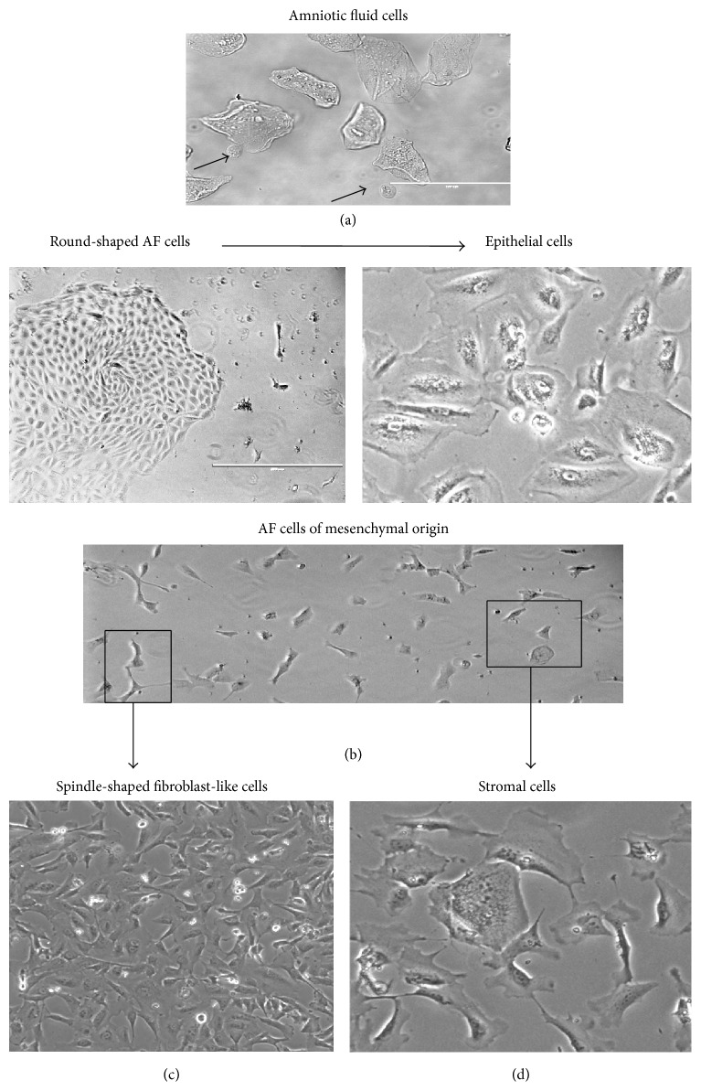
Figure 1.
Morphological characteristics of AF cells. (a) Amniotic fluid cells from amniocentesis sample. (b) The colony appearance of epithelial type at 10–15 days after initiation of the primary culture and the expansion of epithelial cell population at passage 3. (c, d) Mesenchymal-type cells in the primary culture at 10–15 days and after culturing to elongated spindle-shaped or flat “stromal” cell populations at passage 3.

