. 2015 Aug 20;19(5):413–420. doi: 10.4196/kjpp.2015.19.5.413

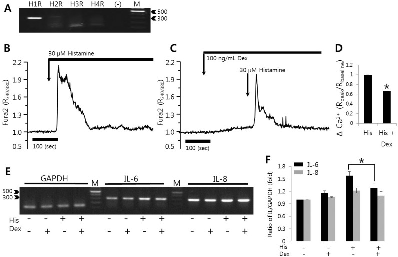
Fig. 5. Histamine-induced [Ca2+]i signals were inhibited by dexmedetomidine in HSG cells. (A) Expression levels of histamine receptor mRNA (H1R to H4R) in HSG cells. (B) Changes in [Ca2+]i induced by 30 µM histamine. (C) Changes in [Ca2+]i after pretreatment with 100 ng/mL dexmedetomidine (Dex) and subsequent treatment with 30 µM histamine (His). (D) Evoked [Ca2+]i (Δ Ca2+) calculated by the peak value of 30 µM histamine stimulation in the presence or absence of 100 ng/mL dexmedetomidine. The upper bars indicate the extracellular solutions applied to the cells and traces were represented with average value. (E, F) After pre-treatment with 100 ng/mL dexmedetomidine for 30 min, HSG cells were stimulated with PBS or with 30 µM histamine for 90 min and then total RNAs were extracted and amplified with primers specific for IL-6, IL-8, and GAPDH. Data are from one of three experimental replicates. IL-6 and IL-8 mRNA expressions were quantified after normalizing to GAPDH levels as a loading control. Results are the mean±SEMs of three independent experiments. (-): no RNA, M: DNA ladder (bp), *p<0.01 was considered statistically significant.

Fig. 5