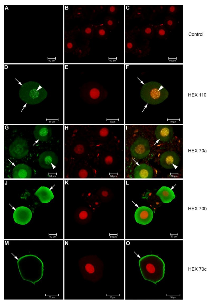
Figure 4.
Confocal microscopy for detection of hexamerins in the fat body oenocytes of pharate pupae (PP phase). Alexa Fluor 488-stained enocytes: preparations (A) without the specific (primary) antibody (control) or with (D) anti-HEX 110, (G) anti-HEX 70a, (J) anti-HEX 70b or (M) anti-HEX 70c for detection of the respective hexamerins (green foci). (B, E, H, K, N) Propidium iodide-stained cell nuclei (red). At the right column, the merged images show: (C) the control without the antibody, (F) HEX 110 and (I) HEX 70a mainly in the nuclei (yellow foci) but also in the cytoplasm (green foci), (L) HEX 70b in the cytoplasm (green foci), (O) HEX 70c at the cytoplasmic periphery (green foci). Arrowheads: nuclear foci of hexamerins. Arrows: hexamerin foci in the cytoplasm.

