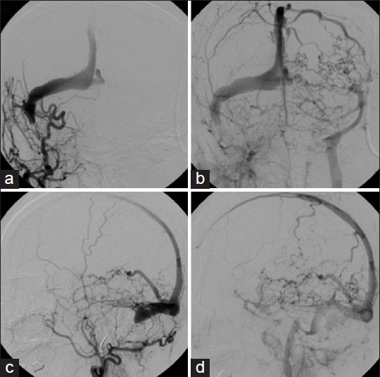
Figure 1.

Preoperative angiogram. Right external carotid artery angiogram (a and c: Early phase, b and d: Late phase) showing a transverse-sigmoid sinus dural arteriovenous fistula with occlusive changes in both the right sigmoid sinus and the left transverse sinus. Marked retrograde venous drainage was directed toward the deep venous system through the straight sinus and toward the cerebral hemisphere via the superior sagittal sinus
