Figure 3.

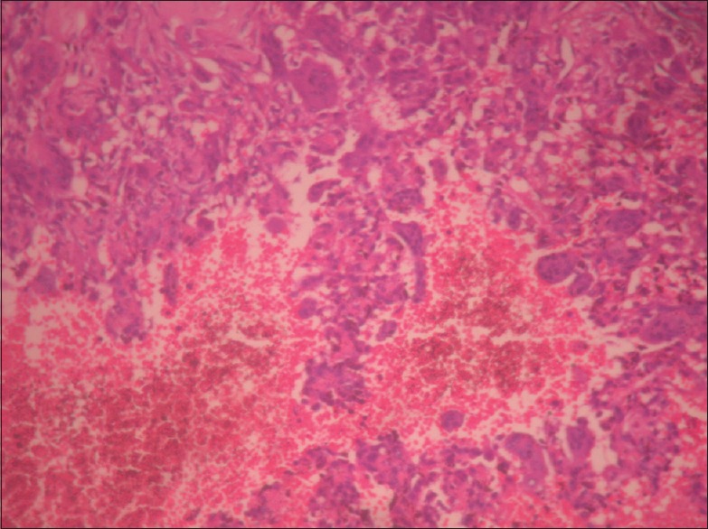
H and E stains showing osteoclast type giant cells having multiple nuclei. Cysts containing haemorrhage and lined by histiocytes and osteoclast giant cells can be noted. There features are suggestive of secondary aneurysmal bone cyst

H and E stains showing osteoclast type giant cells having multiple nuclei. Cysts containing haemorrhage and lined by histiocytes and osteoclast giant cells can be noted. There features are suggestive of secondary aneurysmal bone cyst