
Fig. 2.
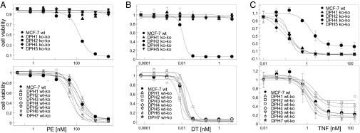
Influence of DPH inactivation on sensitivity to ADP ribosylating toxins and TNF. Dose–responses of cells with inactivation of DPH1, DPH2, DPH4, and DPH5 in comparison with parent MCF7 (bold line), exposed to PE (A), DT (B) or TNF (C). Complete knockouts (Upper) and partial knockous (Lower) are shown. Parent MCF7 (bold line) and partial knockouts show the same sensitivity to PE, DT, and TNF. Cells with complete inactivation of DPH1, DPH2, DPH4, and DPH5 are resistant to PE and DT and hypersensitive to TNF.