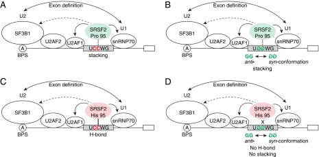
Fig. S7.

A hypothetical model of the functional mechanism of mutSRSF2. Pro-95 in WT SRSF2 (shown as a light green oval) forms a stacking interaction (dashed vertical line) with both the second cytosine in UCCWG sites (A) and the second guanine in UGGWG sites (B). His-95 in mutSRSF2 (shown as a light red oval) may form an H-bond (solid vertical line) with the second cytosine in UCCWG sites (C). An H-bond is generally stronger than a stacking interaction. Because the second guanine in the UGGWG sites is in syn-conformation, it is not possible for His-95 in mutSRSF2 to form an H-bond, yielding weaker binding to UGGWG sites (shown as an X) (D).
