Fig. 3.
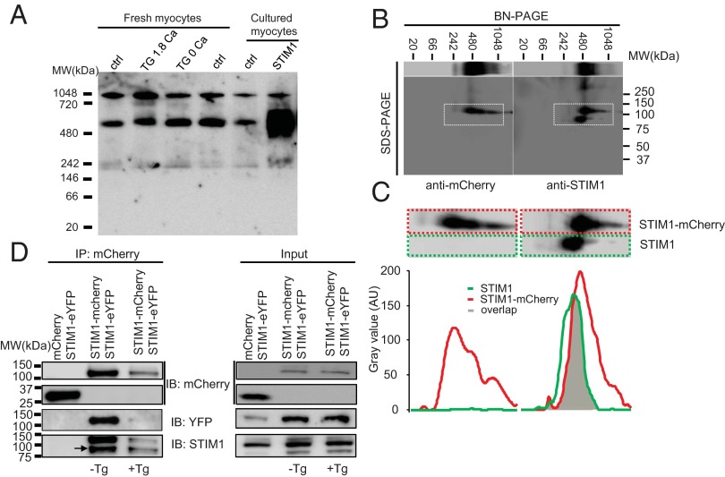
Multimeric properties of STIM1 in rat ventricular myocytes. (A) BN-PAGE shows that endogenous and overexpressed STIM1 forms multimers or protein complex in ventricular myocytes. (B) 2D BN-PAGE and SDS/PAGE show the separation of STIM1 multimer or protein complex using anti-mCherry (to detect STIM1-mCherry, Left panel) and anti-STIM1 (to detect STIM1 and STIM1-mCherry, Right panel). (C) Amplification of the corresponding area marked with the dashed white boxes in B (Upper panel). Band distribution profiles corresponding to the dashed color boxes in the Upper panels show overlap of STIM1 and STIM1-mCherry (Lower panel). Values are presented as arbitrary units (AUs) of the intensity, with background subtracted. (D) Representative (of two independent tests) co-IP shows physical association between STIM1, STIM1-mCherry, and STIM1-eYFP in the absence and presence of Tg (10 µM, 30 min). The lysates from the myocytes overexpressing mCherry and STIM1-eYFP (left lane) or STIM1-mCherry and STIM1-eYFP (middle and right lanes) were immunoprecipitated with anti-mCherry and probed with anti-YFP, anti-mCherry, and anti-STIM1.

