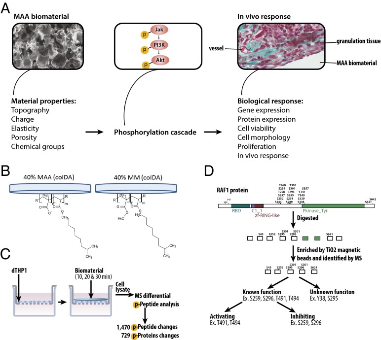
Fig. 1.
Overview of phosphoproteomic method to assess the effects of materials on biological responses. (A) Biological responses (e.g., gene expression, cell proliferation, in vivo host response) are related to material properties (e.g., topography or chemistry) through phosphorylation signaling cascades. The properties of the material govern how proteins from the extracellular milieu adsorb to the surface of the biomaterial and are presented to the cell. These proteins activate the signaling phosphorylation cascades within the cell that drive the biological response to the material. (B) Films containing MAA or MM copolymerized with IDA were cast onto the underside of glass coverslips. (C) Coverslips coated with 40% MAA or 40% MM films were placed on serum-starved dTHP1 cells for 10, 20, or 30 min (with films lying atop cells in a Transwell insert), after which the cells were lysed with urea buffer. The cell lysate was digested with trypsin and the phosphorylated peptides were enriched from the cell lysate by TiO2 magnetic beads and analyzed by MS. (D) Phosphorylated peptides from proteins (RAF-1 is shown as an example) were enriched and identified by MS. Peptides were sorted based on the function of the phosphorylation site. Some proteins had a known function of the phosphorylation site that was further distinguished by literature review; other phosphorylation sites had no known function.

