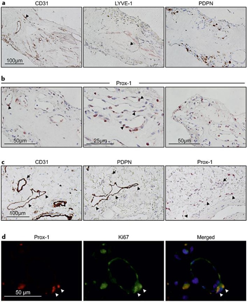
Fig. 2.
LEC markers are expressed in neovascular hemi-RVO tissue specimen. a, b LYVE-1, PDPN, and nuclear transcription factor Prox-1 were used as markers for LECs. LYVE-1 immunoreactivity was observed (arrow head) (a, middle panel); immunoreactivity of PDPN was not found in any vascular structures, only in the extravascular structures (a, right panel), and corresponding pan-endothelial marker CD31 staining is also shown (arrow head) (a, left panel). Prox-1 positivity was observed in vessel-lining cells and also in circulating bone marrow-derived cells (arrow heads) (b). c Immunostaining of CD31, PDPN, and Prox-1 of the skin sample was used as control. Arrows indicate a blood vessel in the corresponding sections, and arrow heads indicate a lymphatic vessel showing positivity for PDPN and Prox-1. d Coimmunostaining of Prox-1 and Ki67 shows active proliferation of Prox-1-positive lumen-lining cells.

