
Figure 2.
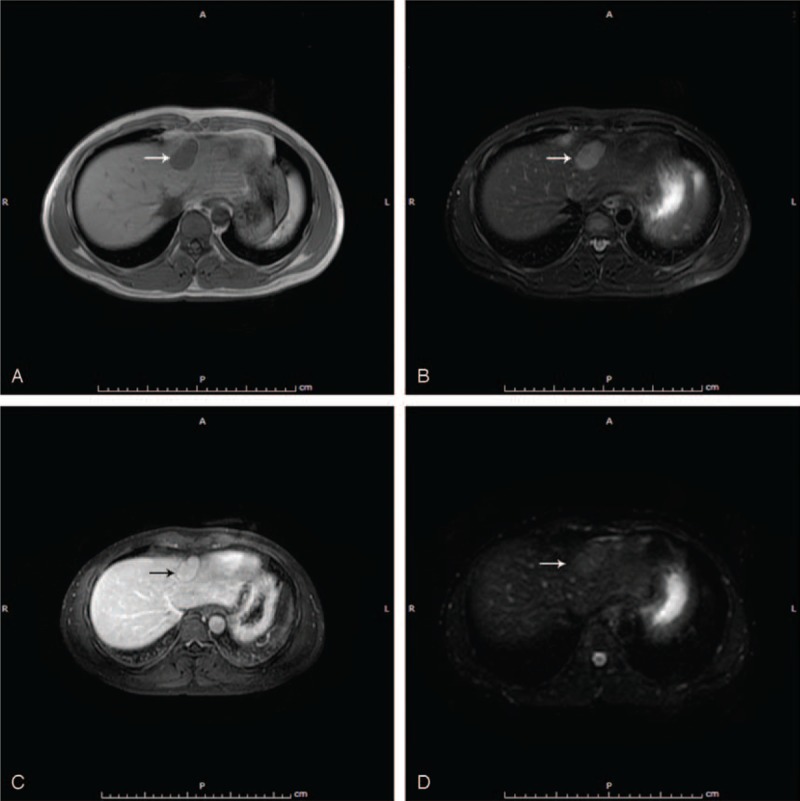
(A) T1-weighted MR image: the lesion is of low signal intensity (white arrow). (B) T2-weighted MR image: the lesion is of high signal intensity (white arrow). (C) Liver acceleration volume acquisition contrast: the lesion is of slight hypointensity (black arrow). (D) DWI: the lesion is of slight hyperintensity (white arrow). DWI = diffusion-weighted imaging, MR = magnetic resonance.