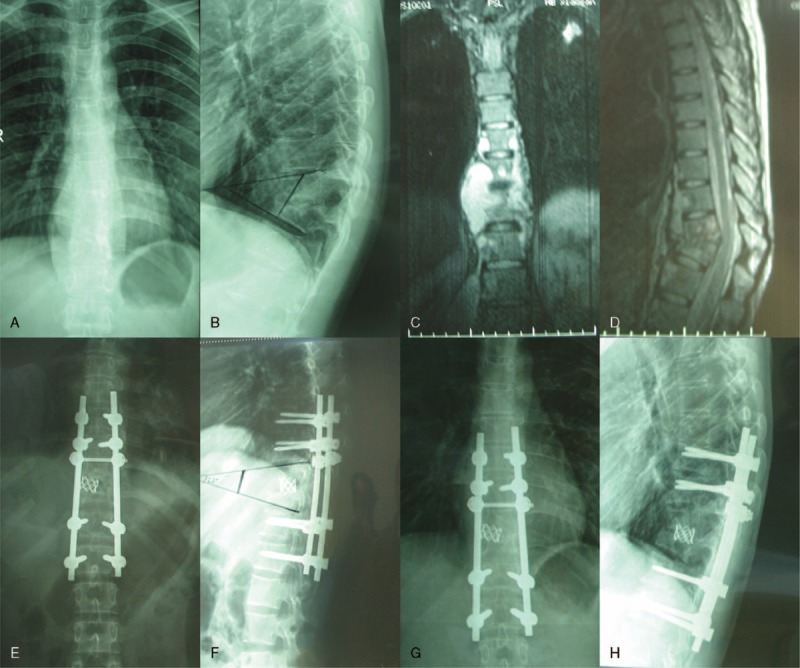
FIGURE 1.

A 25-year-old female patient (a–h), preoperative X-ray (a, b) and MRI (c, d) shows T8, T9 vertebral body bone destruction with cyrtosis, compressed spinal dura mater, and paravertebral abscess. Postoperative 1 week, X-ray (e, f) show vertebral body height corrected with Cobb angle. Postoperative 1 year, X-ray (g, h) showed a good fixed position. MRI = magnetic resonance imaging
