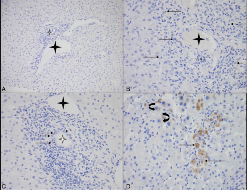
FIGURE 3.

PLA2R immunostaining in normal liver and cases of autoimmune liver diseases (with or without MN). (A) Absence of PLA2R expression in hepatocytes and portal area in normal liver tissue specimen (original magnification ×20). (B) PLA2R immunostaining gave negative results in AIH (control) with portal infiltrate rich in plasmocytes (arrow) with interface hepatitis (original magnification ×40) and (C) in PBC (control) with portal inflammation and bile duct damage (arrow: intraepithelial lymphocyte in bile duct) (original magnification ×40). A similar pattern (no significant PLA2R signal) was observed in patients with autoimmune and/or immunological-related liver disease associated with MN (Table 3). (D) Nonspecific cytoplasmic staining in macrophages (arrow) and hepatocytes (lipofuschines: curved arrow) in patient 5 (original magnification ×40). The interlobular bile duct and portal vein are indicated with white and black stars, respectively. PLA2R = phospholipase A2 receptor.
