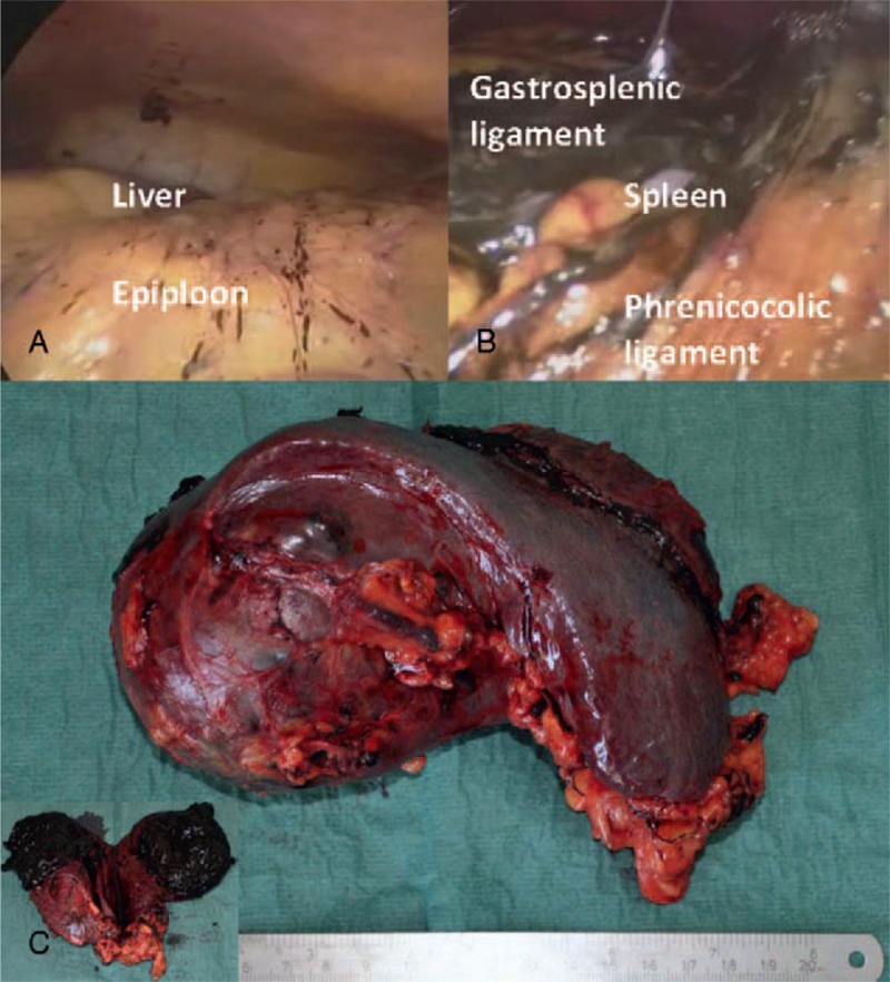
FIGURE 2.

(A) Intraoperative findings of focal peritoneal melanosis on the epiploon and on the peritoneal surface of the right diaphragmatic peritoneum; (B) massive amount of peritoneal melanosis around the spleen, on gastrosplenic and phrenicocolic ligaments; (C) Surgical specimen showing a large brownish irregular round mass that protrudes above the surface at the upper pole of the spleen; a cut surface shows the dishomogeneous appearance and the intense dark brown color of the lesion, with soft solid component and areas of colliquation.
