
Figure 1.
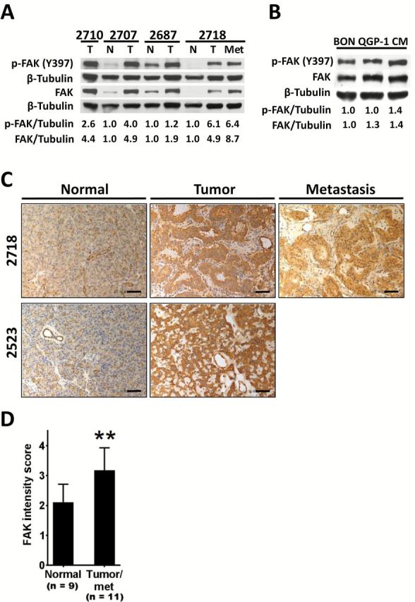
Focal adhesion kinase (FAK) expression in pancreatic neuroendocrine tumors (PanNETs). A) Immunoblot analysis of Y397 phospho- and total FAK in PanNET patient tumors (T) and/or matched metastatic sample (Met) as compared with matched normal pancreas (N). Numbers (2710, etc.) indicate samples obtained from the same patient. For densitometric analysis, results were normalized to tubulin and are expressed as fold induction over matched normal sample. For the unmatched tumor sample, results are expressed as fold induction over the mean of the other three normal samples. B) Immunoblot analysis of phospho- (Y397) and total FAK in human PanNET cell lines. Tubulin is shown as a loading control, and densitometric analyses are expressed as fold induction over BON cells. C) Representative images of FAK immunohistochemistry staining in human PanNET samples (scale bar = 200 µm). D) Analysis of FAK immunohistochemistry staining intensity in nine normal samples and 11 matched PanNET tumor samples (including one unmatched tumor and one matched metastasis sample) as determined by a masked pathologist using a two-tailed Student’s t test (**P < .01). Error bars represent standard deviation of the mean. FAK = focal adhesion kinase.