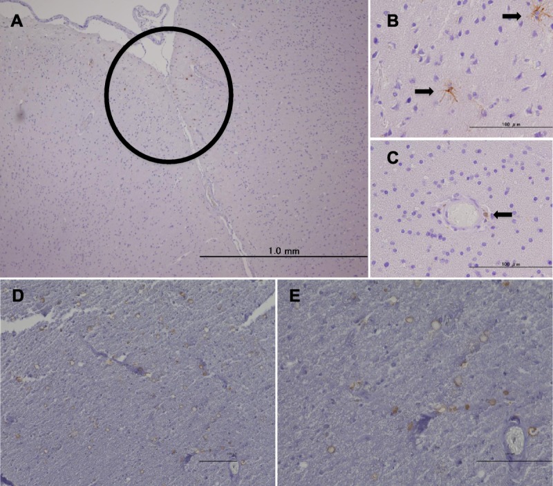
FIGURE 1.

Immunohistochemistry for HLA-A2 with hematoxylin counterstaining. (A) HLA-A2–positive cells accumulate in the cerebral cortex (brown cells within the circle). (B) HLA-A2–positive cells with ramified morphology (arrows). (C) A round HLA-A2–positive cell around a vessel (arrow). HLA-A2–positive cells with a round or ring morphology in the C5 lesion of the spinal cord at lower magnification (D) and at higher magnification (E). Scale bar = 100 μm.
