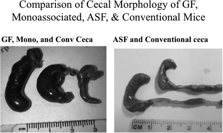
Figure 1.
Morphological features of the ceca from gnotobiotic and conventional mice. Left panel: Representative images of the cecum excised from a germfree (left), a monoassociated (center), or a conventional (right) C3H/HeN mouse. Right panel: Representative images of the cecum excised from an ASF (left) or conventional (right) C3H/HeN mouse.

