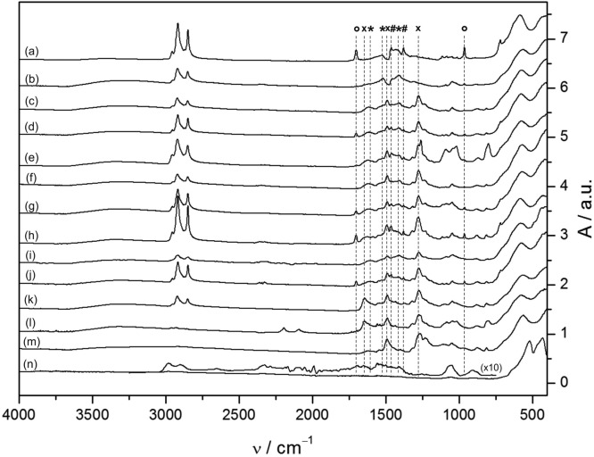
Figure 2.
ATR–FTIR spectra of various 3.5 nm magnetite nanoparticle preparations: (a) as-synthesized SPION containing excess physisorbed OA, (b) purified SPION with a monolayer of chemisorbed oleate, (c–j) mixed dispersant OA/P-NDA–SPION and (l–m) post-coated, pure nitrocatechol-ligand-capped SPIONs. SPIONs were purified by the following methods: (b) pre-extraction in hot MeOH containing 1 mM OA as a stabilizer, (c) hot MeOH extraction, (d) cold MeOH extraction, (e) syringe filtration (PTFE), (f) surfactant addition (CTAB), (g) silica column chromatography (THF/MeOH = 4:1), (h) ligand saturated chromatography, (i) repeated MeOH/n-hexane (1:1) recrystallization, (j) 2,6-lutidine as a solvent, (k) post-coated P-NDA–SPION, (l) post-coated d31P-NDA–SPION, (m) post-coated NDA–SPION, and (n) SPION with combusted shell (post-TGA) with 10× scaled inset of residual absorptions. Peaks corresponding to physisorbed and chemisorbed OA are indicated by circles and asterisks, respectively, while crosses depict bands related to nitrocatechol ligands. The post-coated P-NDA (k), d31P-NDA (l), and NDA particles (m) are the only particles that demonstrate an absence of the characteristic OA bands and, therefore, complete ligand replacement.

