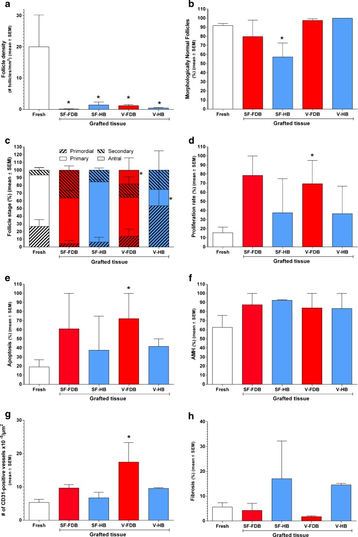
Fig. 2.
Impact of the cryopreservation technique and vascular bed on different ovary and follicle characteristics after grafting. Follicle density (a), morphologically normal follicles (MNFs; b), follicle stage (c), proliferation rate (d), apoptosis rate (e), follicle functionality (expressed as a percentage of AMH-positive follicles; f), vascularization (expressed as a percentage of CD31-positive vessels ×105/μm2; g), and fibrosis (expressed as a percentage of area stained with Masson’s trichrome; h) are shown in the figure. Fresh ovarian tissue (fixed immediately, open bar), slow-frozen (SF) and grafted ovarian tissue, and vitrified (V) and grafted ovarian tissue were used. SF and V ovarian tissue were grafted to a freshly decorticated bed (FDB, red bars) or healed bed (HB, blue bars). Mean ± SEM are depicted on the graph. Differences were considered significant when q values were <0.2. Asterisk vs fresh tissue: q < 0.2

