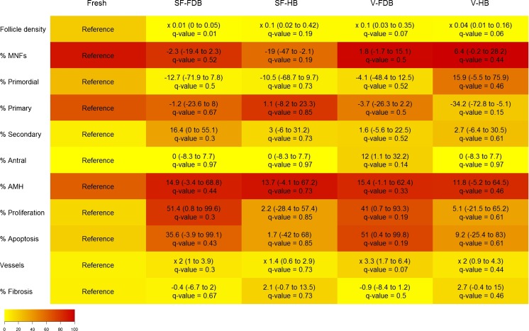
Fig. 3.
Impact of the cryopreservation technique and vascular bed on different ovary and follicle characteristics after grafting (heatmap). Increased (positive values) or decreased (negatives values) effects of different treatments applied to ovarian tissue compared to the fresh tissue used as a reference are shown on the graph. Parameters studied were follicle density, morphologically normal follicles (MNFs), follicle stage, proliferation rate, apoptosis rate, follicle functionality (expressed as a percentage of AMH-positive follicles), vascularization (expressed as a percentage of CD31-positive vessels ×105/μm2), and fibrosis (expressed as a percentage of area stained with Masson’s trichrome). Fresh ovarian tissue (fixed immediately, fresh), slow-frozen (SF) and grafted ovarian tissue, and vitrified (V) and grafted ovarian tissue were investigated by transplanting to a freshly decorticated bed (FDB) or healed bed (HB). The cell color intensity (more reddish or more yellowish) reflects the average value of each parameter in each group. 95 % CI and q values are indicated on the graph. Differences were considered significant when q values were <0.20

