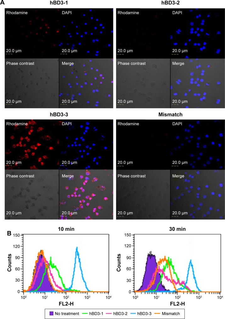
Figure 2.
Intracellular translocation of hBD3-3 in vitro.
Notes: (A) Cellular localization of rhodamine-labeled peptide fragments in RAW 264.7 cells. Cells (1×10⁴) were incubated for 10 minutes in medium containing the rhodamine-labeled peptides (50 μM) (original magnification 40×). (B) FACS analysis of cells treated with rhodamine-labeled peptides. Cells (1×10⁶) were incubated for 10 minutes and 30 minutes in medium containing the rhodamine-labeled peptides (50 μM).
Abbreviations: hBD3, human beta-defensin 3; FACS, fluorescence-activated cell sorting; DAPI, 4′,6-diamidino-2-phenylindole; min, minutes.

