Figure 3.
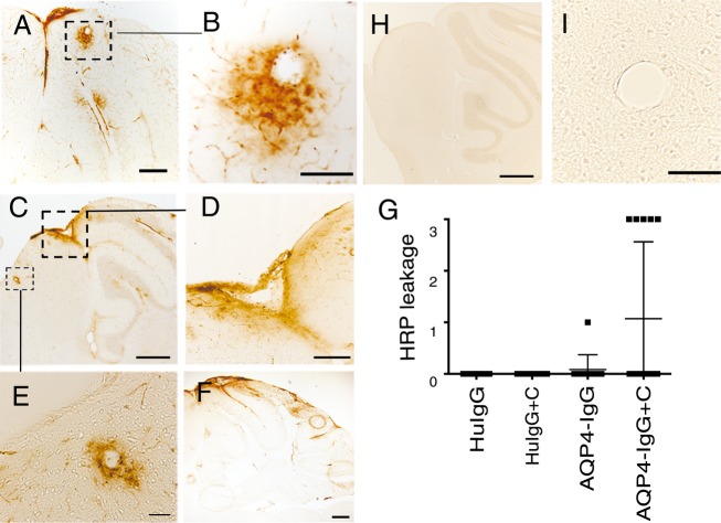
BBB breakdown associates with CSF-derived AQP4-IgG + complement and perivascular astrocyte pathology. Micrographs show sagittal sections of the brain of animals injected with AQP4-IgG + complement. BBB breakdown was visualized by leakage of intravenously injected HRP as tracer (brown). (A–F) Micrographs show HRP leakage into the brain parenchyma in cerebellum and midbrain, (D) shows a magnified view of an area in (C) in midbrain. (B and E) show magnified views of perivascular HRP leakage shown in (A and C) respectively, as wide halos around blood vessels. Such pathology was not seen in controls mice (H and I). (G) Quantitation of HRP leakage into the brain parenchyma in animals injected with AQP4-IgG + complement (n = 14), AQP4-IgG alone (n = 12), control HuIgG (n = 10), control HuIgG + complement (n = 13), expressed on a scale of 0–3. Data are presented as mean ± SD. Data indicate BBB breakdown in AQP4-IgG + complement-treated mice. Bar 50 μm (B and D), 20 μm (E), 100 μm (A and F), 200 μm (C, F, and H). BBB, blood-brain barrier; CSF, cerebrospinal fluid; HRP, horseradish peroxidase.

