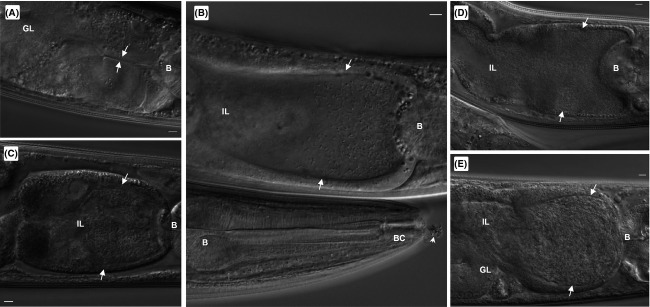
Figure 1.

Legionella pneumophila colonizes the Caenorhabditis elegans intestinal lumen. Representative DIC images of N2 nematodes fed live Lp02 over time: (A) 2 days p.i.; (B) 3 days p.i.; (C) 4 days p.i.; (D) 6 days p.i.; and (E) 8 days p.i. Note also that the integrity of the intestinal lumen remains intact despite the progressive distension of the intestinal lumen due to the accumulation of colonized bacteria featuring typical rod-shaped morphology (white arrows). Anatomical features indicated in the microscopic images include the terminal bulb (B), buccal cavity (BC), intestinal lumen (IL) and the gonadal loop (GL). Scale bar represents 5 μm in all panels.
