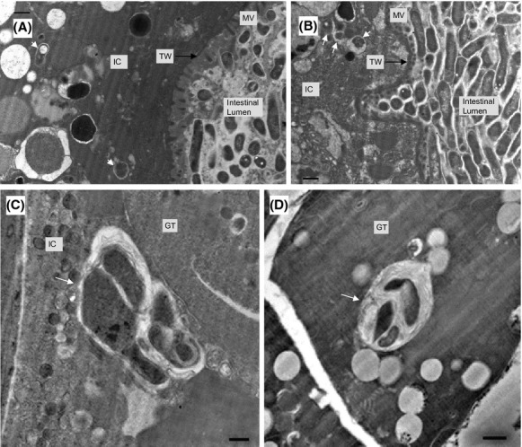
Figure 8.

Morphological differentiation of internalized Legionella pneumophila bacterial forms. Representative transmission electron microscopy images were obtained from samples of nematodes fed L. pneumophila Lp02 for 6 days. (A and B) Internalized bacterial forms (white arrow) within intestinal cells (IC) with microvilli (MV) on the apical surface supported by the terminal web (TW). Note that the bacterial membrane is more defined due to progressive formation of multiple layers. (C) Irregular- shaped transitional bacterial forms (white arrow) inside an IC next to the gonadal tissue (GT) (i.e., gonad arm). (D) Presence of a well-defined Legionella-containing vacuoles, containing irregular-shaped transitional forms and membrane whorls (indicated by white arrow), in a GT cell of the gonad arm. Membrane whorls most likely represent eukaryotic membrane fragments. Scale bar is 500 nm for all panels.
