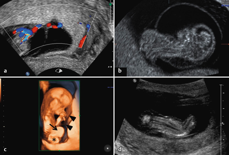
Fig. 2 a.

to d Ultrasound findings in the body stalk anomaly. a Short umbilical cord. b Partial extraamniotic position of the fetus: the upper body is surrounded by the circular depicted amnion, while the lower body lies outside of the amnion. c 3D surface image of the same fetus as in b. A large abdominal wall defect is visible with herniation of liver (→) and intestine (*). The amnion can also be seen (▸) – it extends up to the abdominal wall defect. d Scoliosis in a different fetus with body stalk anomaly.
