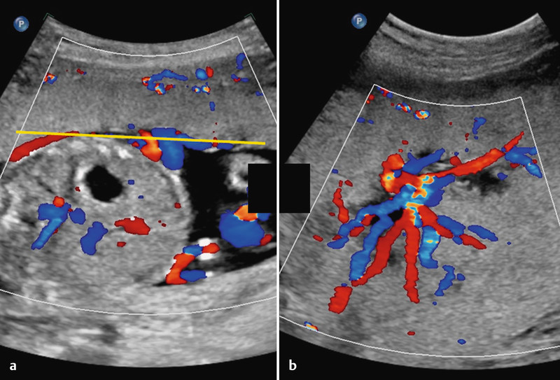
Fig. 3 a.

and b Normal umbilical cord insertion. a Demonstration of the cord insertion and a number of chorionic plate vessels. Depending on fetal lie visualisation may be difficult even if the placenta is anterior. For further differentiation it may be helpful to examine the placenta tangentially as in b. The star-like pattern of the chorionic plate vessels as they approach the cord insertion is seen. Placental tissue surrounds them. The yellow line in a represents the level and orientation of the image in b.
