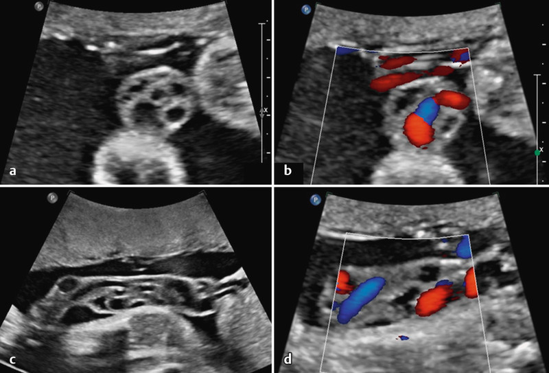
Fig. 5 a.

to d Cystic segment of UC in cross section (a und b), and in longitudinal section (c und d). Cysts can be differentiated from perfused vessels using Doppler ultrasound (b und d). Despite the cystsʼ segmental occurrence and irregular shape, both characteristic of pseudocysts, differentiation is only possible on histology. This fetus also had agenesis of the septum pellucidum, schizencephaly and atrioventricular valve incompetence.
