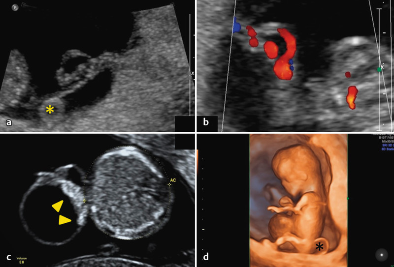
Fig. 6 a.

to d a and b Demonstration of an UC cyst in a central cord segment in the first trimester; c and d Fetus with a large UC cyst located at the fetal cord insertion. The echogenic appearance of the cyst margin nearest the abdominal wall (▸) raises the suspicion of an abdominal wall defect. An omphalocele was subsequently diagnosed. Caption: yolk sac (*).
