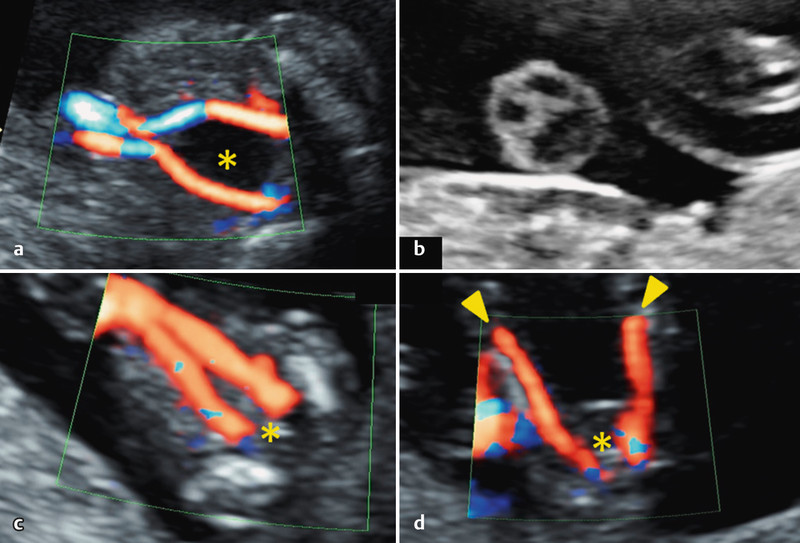
Fig. 7 a.

to d a Perivesical course of the UAs right up to the fetal cord insertion. b Cross section of a normal umbilical cord. The three vessel lumens appear as a stylized Mickey Mouse. c Demonstration of two UAs in their perivesical course in the first trimester up to the fetal cord insertion. d Due to their close proximity in the first trimester, the UAs may be confused with the femoral arteries (▸). Caption: urinary bladder (*).
