
Fig. 9 a.
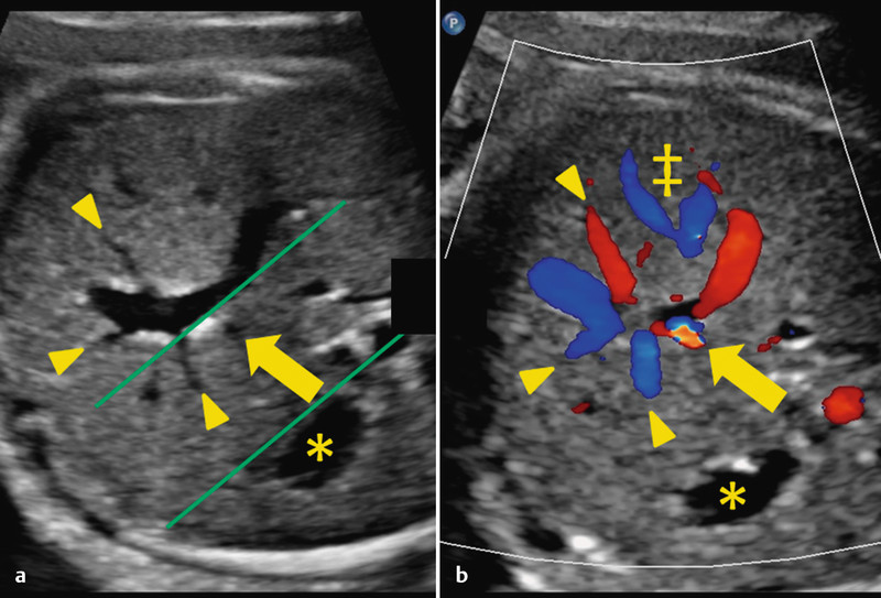
and b Normal anatomy: intraabdominal course of the UV and union with portal system. Green lines in section a show the tangential course of the arching vessel with respect to the fetal stomach (*). The left portal vein branch (▸) and ductus venosus (→) are visible. In section b a colour change allows identification of the ductus venosus (“aliasing” caused by flow acceleration). Caption: hepatic veins (‡).