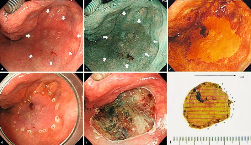
Fig. 1 a.

Reddish flat elevated lesion located in the right pyriform sinus of the hypopharynx. b A well demarcated brownish area demonstrated using narrow-band imaging. c Chromoendoscopy using Lugol staining demonstrated the unstained lesion. d Marking dots around the lesion. e Endoscopic submucosal dissection (ESD) ulceration made by en bloc resection without any complications. f Macroscopic image of the resected specimen. Histopathological examination revealed squamous cell carcinoma with subepithelial invasion.
