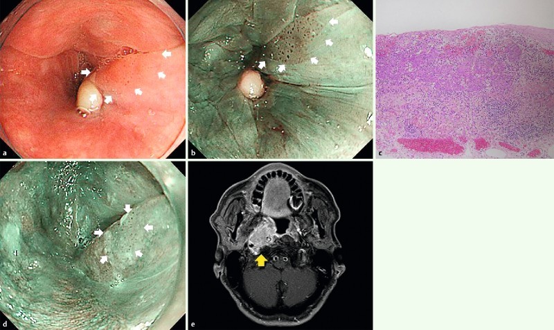
Fig. 5.

Endoscopic and CT findings of LNM case-3. a, b White light and narrow-band imaging showed 0-IIa in the left pyriform sinus of the hypopharynx. EMR was performed for this lesion. c Histopathological examination revealed slight subepithelial invasive cancer, with no lymphovascular invasion, horizontal margin positive, vertical margin negative. d Twelve months after ER, local recurrence was detected and argon plasma coagulation (APC) was performed on this lesion. e Twelve months after APC, CT showed lateral retropharyngeal lymph node enlargement.
