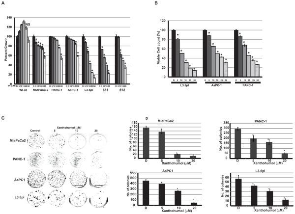
Figure 1.
Effect of XN on pancreatic cancer cellular proliferation. A. Human pancreatic cancer cell lines and human normal lung fibroblast cells (WI-38) were treated with XN at indicated doses for 24 hours and cytotoxicity was measured using MTT assay (n=3; p=0.05 at 20 μM concentrations for all cancer cell lines except WI-38; not significant compared to control). B. Viable cells were counted after 48hr of XN treatment using trypan blue staining. (n=3; p<0.05 for all cancer cell lines). C. Cells were treated with XN up to 20μM for 4 days and cell viability was measured by colony formation assay. D. Bar graph shows the number of colonies formed after XN treatment (n=3; *p>0.05 in all treatment; NS, not significant).

