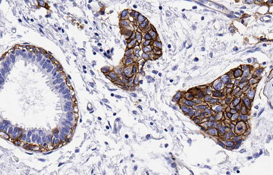
Figure 3. Altered localization of integrin β4 expression in invasive breast carcinoma.
In a dilated duct with benign columnar cell change (left), integrin β4 is expressed in myoepithelial cells surrounding the duct, but is absent in luminal cells. Adjacent nests of invading carcinoma cells (right) display elevated expression of the integrin β4. Immunohistochemical staining was performed using the 439-9B antibody as previously described (72). Brown staining represents positive expression of the integrin β4.

