
Figure 4.
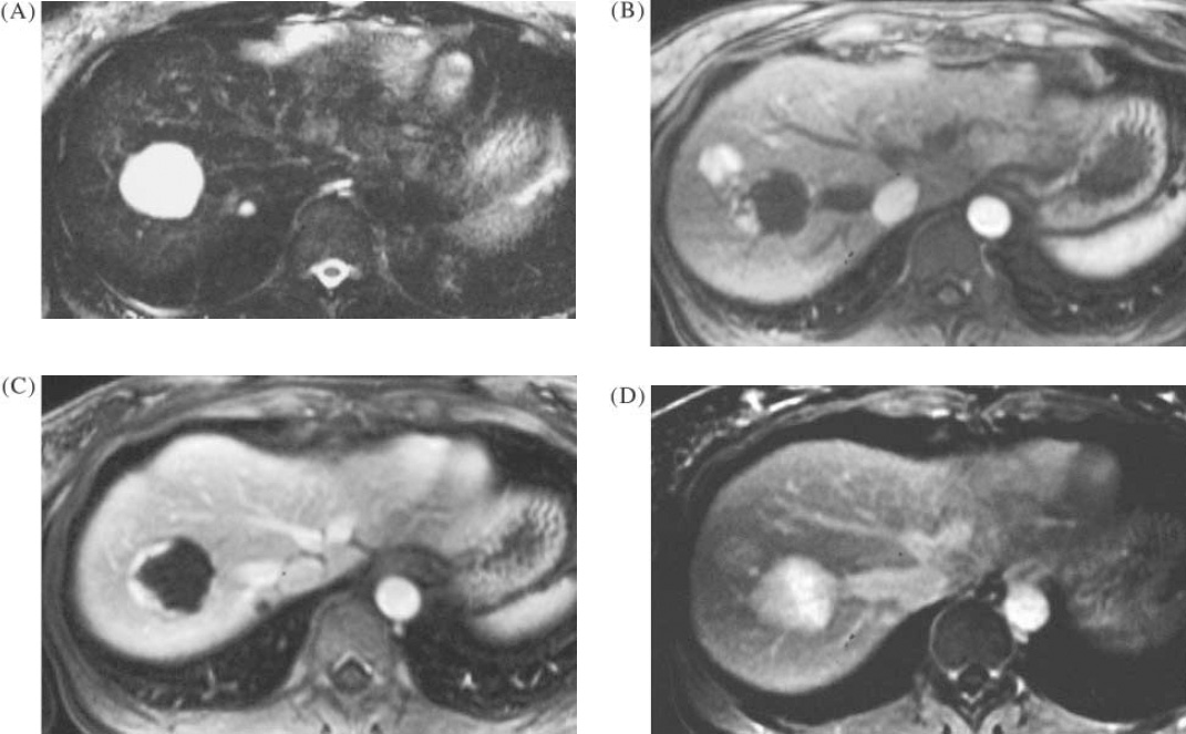
Benign liver lesions in a patient with colorectal cancer. (A) TSE T2 image showing 3 cm lesion which was indeterminate on ultrasound, also a second lesion of 7 mm. (B) and (C) T1-weighted image with gadolinium enhancement, arterial phases, showing typical peripheral nodular enhancement in the larger lesion but also an additional hypervascular lesion. Delayed post-gadolinium T1 image showing complete infilling of the 3 cm and 7 mm lesions, typical for haemangioma, and a faint persistent stain in the hypervascular lesion, suggestive of focal nodular hyperplasia. The FNH lesion was undetected on T2-weighted MRI, and also on ultrasound.