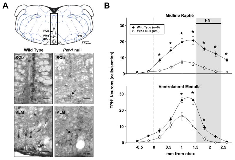
Figure 1.
A) A representative sketch (upper panel) of a section ~ −6.5mm from Bregma with an overlaying 0.5 mm rectangle centered on the midline, and extending to dorsal and ventral surfaces. This rectangle represents the midline count region, and includes the midline raphé nuclei (raphé obscurus, pallidus, and magnus). All other TPH+ neurons were included in the VLM region counts. Grayscale images of WT (lower left panels) and Pet-1 null mice (lower right panels) transverse medullary sections (20 μm) immunostained for TPH. Note that TPH+ 5-HT neurons were fewer in number, and generally lighter in staining intensity in Pet-1 null (arrows) compared to control mice in the midline and ventrolateral medullary (VLM) regions. B) The number of TPH+ neurons in WT (n=9) and Pet-1 null (n=9) brainstems along the midline (upper panel) and VLM regions (binned every 0.5 mm) are plotted using the obex (dotted line) as the reference point. Note that there were significantly fewer TPH+ 5-HT neurons in the midline (-73.2%) and VLM (-50.9%) regions in Pet-1 null mice.

